- Noticias / Researchers achieve the highest resolution in fluorescence microscopy
Scientific advance
Researchers achieve the highest resolution in fluorescence microscopy
F.Stefani, part of an international team led by S.Hell, Nobel Prize in Chemistry, developed a method to achieve an original resolution in living cells.
Compartir en
redes sociales
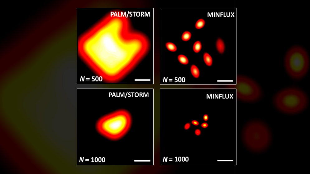
Fernando Stefani, deputy director at the Centro de Investigaciones en Bionanociencias (CIBION) of CONICET and independent researcher of the Council, participated in a study in which the scientists managed to go beyond the limit resolution of the fluorescence microscopes called super-resolution ones. The researchers developed a new method called MINFLUX (for its minimal use of photons), which was presented in Science and allows experts to see the details of a nanometer (one millionth of a millimeter), that is to say, 10 million times smaller than a centimeter.
“Fluorescence molecules emit a limited number of photons. This restricts the space resolution that can be reached with the super-resolution microscopy. This new MINFLUX method combines several concepts of high resolution fluorescence microscopy in a new way, squeezes all the information of each fluorescence photon in order to determine the position of one molecule. For this reason, it enables us to get a new resolution level, reaching the same dimensions of only one molecule, one nanometer”, Stefani explains.
One of the main authors of the study is a young Argentine, Francisco Balzarotti, who works at the Depart Department of NanoBiophotonics of the Max Planck Institute of Biophysical Chemistry in Germany, and is one former member of the Facultad de Ciencias Exactas y Naturales of the UBA (FCEN-UBA).
“Stefan Hell’s collaboration began in 2011 when we obtained one Partner Group subsidy from the Max Planck Society. Francisco was the first one to be involved in this study, travelled to Germany as a postdoctoral fellow and made a great job. After that, the project was developed elaborately and more researchers from Germany and Sweden joined”, Stefani comments.
The scientists believe that this new technology will allow this type of technology to compete with the electronic microscopy, which provides nanometric spatial resolution but imposes adverse conditions to observe living organisms. “In contrast to electronic microscopy, MINFLUX can be implemented in a conventional laboratory, does not require high technology and the fact that they use light instead of energetic electrons is convenient to observe biological samples.” In this article, scientists show the MINFLUX application to the monitoring of proteins inside a bacteria, a living Escherichia Coli (E. Coli).
This new device is available for Stefan Hell’s group, where Balazarotti works. The CIBION is at the Polo Científico Tecnológico, which would be the second laboratory in the world to have this technology. “We have the knowledge and the capacity to implement MINFLUX and there is one fellow working on this project. We are going to seize this strategic opportunity and apply this new technology in local high impact research.”
“This potential is great. The super-resolution techniques have not been established and this fact leads to a new outlook. We have reached the highest resolution level which has a physical sense for an optical technique, that is to say, of the same size of the source of light, one molecule”, the researcher concludes.
By María Bocconi
About the study:
Francisco Balzarotti. Max Planck Institute of Biochemistry, Germany.
Yvan Eilers. Max Planck Institute of Biochemistry, Germany.
Klaus C. Gwosch. Max Planck Institute of Biochemistry, Germany.
Arvid H. Gynnå, Science for Life Laboratory, Uppsala University, Sweden.
Volker Westphal. Max Planck Institute of Biochemistry, Germany.
Fernando D. Stefani. CIBION and Departamento de Física. FCEN. UBA.
Johan Elf. Science for Life Laboratory, Uppsala University, Sweden.
Stefan W. Hell. Max Planck Institute of Biochemistry, Germany.